Lipoma or Cancer? How to Check Your Dog’s Lumps at Home

Finding an unexpected lump or bump on your canine companion is a moment that can send any pet owner’s heart racing. The mind immediately jumps to the worst-case scenario: cancer. While this fear is valid and understandable, it’s important to approach the situation with a calm and methodical mindset. The vast majority of lumps found on dogs are benign, with the most common being lipomas—harmless fatty tumors. However, the possibility of malignancy means that no new growth should ever be ignored.
This guide is designed to serve as your first resource. It is not a substitute for professional veterinary diagnosis, which is the only way to definitively determine the nature of a lump. Instead, our goal is to empower you with the knowledge to perform a safe and thorough preliminary examination at home. By learning what to look for, how to track changes, and when to seek immediate professional help, you can transform your anxiety into proactive care. You will learn the key characteristics that differentiate a common lipoma from a more concerning growth and understand the critical red flags that warrant an urgent call to your veterinarian. Let’s walk through this process together, ensuring you are the most effective advocate for your dog’s long-term health and well-being.
Understanding Common Lumps and Bumps in Dogs
Understanding Common Lumps and Bumps in Dogs
Before you can assess a lump, it’s helpful to understand the landscape of what’s possible. Lumps and bumps, medically known as masses or tumors, can appear on or under the skin for various reasons. They are broadly categorized as either benign (non-cancerous) or malignant (cancerous).
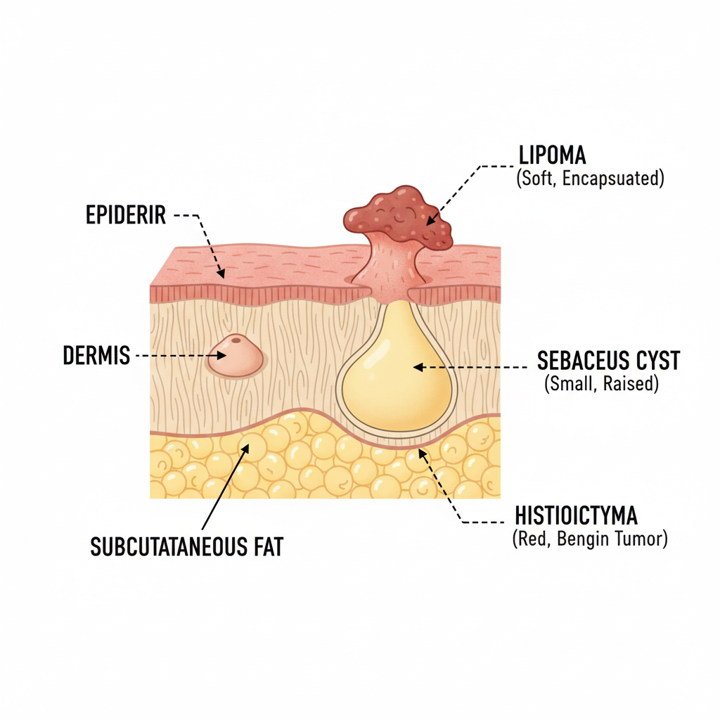
Common Benign Growths
These are non-cancerous and typically do not spread to other parts of the body. While usually harmless, they can sometimes grow large enough to cause discomfort or impede movement, potentially requiring removal.
- Lipomas: By far the most common tumor found in dogs, especially in middle-aged to senior, and often overweight, individuals. These are soft, movable, well-defined masses made of fat cells that sit just under the skin. They rarely cause problems unless their location or size interferes with mobility.
- Sebaceous Cysts: These are essentially blocked oil glands that form a raised, round bump. They can sometimes rupture, releasing a thick, white, or cheesy substance. While generally harmless, they can become infected.
- Histiocytomas: Often called ‘button tumors,’ these are small, red, hairless bumps that typically appear suddenly on young dogs (under three years old). They are benign and often regress and disappear on their own within a few months.
- Skin Tags (Acrochordons): These are fleshy, stalk-like growths that are common in older dogs. They are harmless but can get caught on collars or grooming tools, causing bleeding or irritation.
- Warts (Papillomas): Caused by a virus, these typically appear in or around the mouth of younger dogs but can be found on the skin of older dogs as well. They often have a cauliflower-like appearance and usually resolve without treatment.
Potentially Malignant Growths
These are cancerous tumors that have the potential to grow aggressively and spread (metastasize) to other parts of the body, such as the lymph nodes and internal organs. Early detection is paramount for successful treatment.
- Mast Cell Tumors: These are the most common skin cancer in dogs. They can vary widely in appearance, from small, raised bumps that look like insect bites to larger, ulcerated masses. They are known as ‘the great pretenders’ and require immediate veterinary attention.
- Melanomas: While often associated with pigmented (dark) growths, melanomas can also be non-pigmented. They are commonly found on the mouth, toes, or eyes and can be highly aggressive.
- Soft Tissue Sarcomas: This is a broad category of tumors that arise from connective tissues. They often feel like firm, lumpy masses deep within or under the skin and can be locally invasive.
- Squamous Cell Carcinomas: These tumors often appear as firm, raised, and sometimes ulcerated lesions, frequently on areas with less fur and more sun exposure, like the nose, ears, and belly.
The following table provides a general comparison, but remember these are only typical characteristics. Exceptions are common, reinforcing the need for a professional diagnosis.
| Characteristic | Typical Lipoma | Potentially Malignant Tumor |
|---|---|---|
| Consistency | Soft, pliable, slightly squishy | Often firm, hard, or dense; can have mixed textures |
| Mobility | Easily movable under the skin; not attached to underlying muscle | Often feels fixed or attached to the tissue underneath |
| Growth Rate | Very slow-growing; may remain the same size for months or years | Often grows rapidly or changes noticeably in a few weeks or months |
| Shape & Surface | Typically round or oval with smooth, well-defined edges | Often irregularly shaped with poorly defined borders; may have an ulcerated or raw surface |
| Dog’s Reaction | Usually not painful or sensitive to the touch | May be painful, tender, or itchy; the dog may lick or chew at the area |
A Step-by-Step Guide to Performing a Home Lump Check
A Step-by-Step Guide to Performing a Home Lump Check
Regularly checking your dog for lumps is as important as any other grooming or care routine. Aim to perform a thorough check at least once a month. Choose a time when your dog is calm and relaxed, perhaps while you are cuddling on the couch.
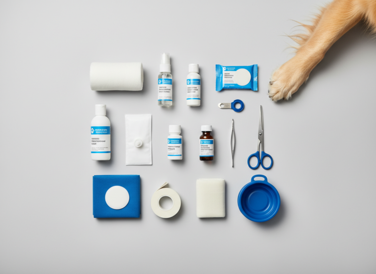
- Prepare Your Space and Your Dog: Find a quiet, well-lit area. Have a notepad and a flexible measuring tape or calipers handy. Start with a gentle petting session to relax your dog completely. Speak in a calm, soothing voice throughout the process.
- Visual Inspection First: Before you start touching, give your dog a full visual scan. Look for anything unusual, such as raised areas, patches of hair loss, or discolored skin. Be sure to check less obvious areas like between the toes, under the tail, in the armpits, and around the ears and mouth.
- The Palpation Process: Using the flats of your fingers (not the tips), gently run your hands over every inch of your dog’s body. Start at the head and work your way methodically to the tail. Apply gentle but firm pressure, enough to feel what’s underneath the skin. Pay attention to the symmetry—if you feel something on one side, check the corresponding spot on the other. Ribs, hip bones, and lymph nodes can sometimes be mistaken for lumps, so familiarize yourself with your dog’s normal anatomy.
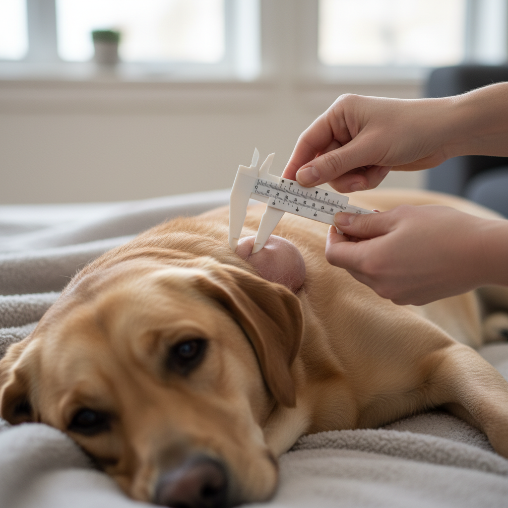
- Examine the Lump’s Characteristics: When you find a lump, do not panic. Calmly and gently assess its features. Lightly pinch the skin next to the lump and try to move it back and forth.
- Size: Use your measuring tape or calipers to get an accurate measurement of its diameter. Estimate its height as well.
- Shape: Is it round, oval, or irregular? Are the borders smooth and easy to define, or do they blend into the surrounding tissue?
- Consistency: Gently press on it. Is it soft and squishy like a water balloon (typical of a lipoma), or is it hard and unyielding like a rock?
- Mobility: Does it move freely with the skin, or does it feel anchored to the muscle or bone underneath?
- Surface: Is the overlying skin and fur normal, or is it red, irritated, ulcerated, or hairless?
- Check for Pain and Other Lumps: Note if your dog shows any signs of pain when you touch the lump, such as flinching, yelping, or trying to move away. After you’ve finished examining the first lump, continue your check over the rest of the body. It’s not uncommon for dogs, especially seniors, to have multiple benign lumps.
Expert Tip: Never squeeze, poke, or attempt to ‘pop’ a lump. Doing so can cause pain, inflammation, infection, and in the case of a malignant tumor, potentially spread cancerous cells. Your role is to observe and document, not to diagnose or treat.
Documenting Your Findings: The ‘Lump Map’
Documenting Your Findings: The ‘Lump Map’
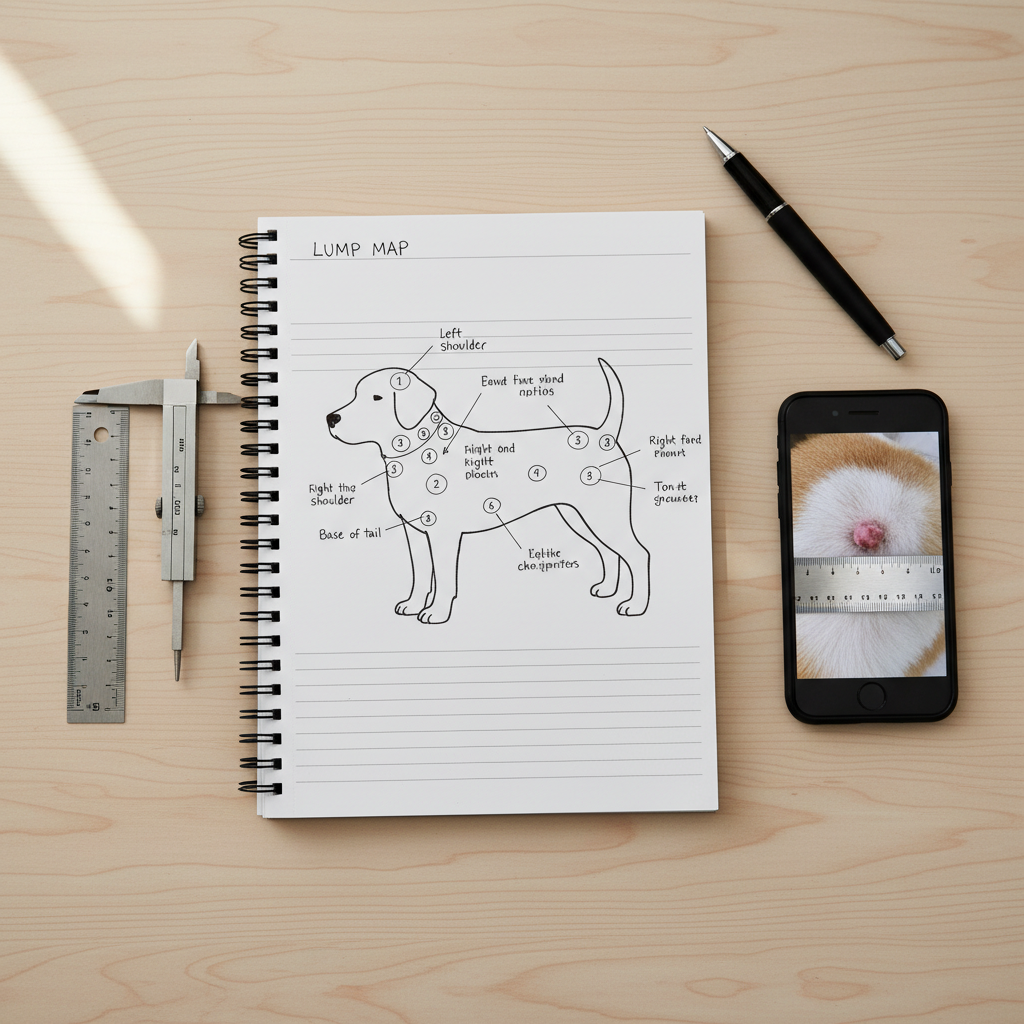
Your observations are only as good as your ability to recall them accurately for your veterinarian. Human memory is fallible, especially when stressed. Creating a ‘lump map’ or a health journal is the single most effective way to track changes over time. This documentation will be invaluable to your vet, as the history and behavior of a mass are critical diagnostic clues.
What to Record
For every lump you find, create a new entry in a dedicated notebook or a digital file. Include the following details:
- Date of Discovery: The exact date you first noticed the lump.
- Location: Be as specific as possible. Instead of ‘on his side,’ write ‘on the right side of the chest, 5 inches behind the armpit.’ You can even print a simple dog body diagram from the internet and mark the location directly on it.
- Size & Shape: Record the measurements (length, width, and estimated height) in millimeters or inches. Note if it’s round, oval, or irregular.
- Appearance: Describe the color of the lump and the overlying skin. Note if it is haired, hairless, ulcerated, scabby, or bleeding.
- Consistency & Mobility: Write down your assessment—’soft and movable’ or ‘hard and fixed.’
- Photographs: Take clear, well-lit photos of the lump from a few different angles. Include a small ruler in the photo for scale. This provides an objective visual record that is superior to memory alone.
The Importance of Monitoring
Once you have your initial record, set a reminder to re-check and update it every two to four weeks. If you notice any change—in size, shape, color, or texture—contact your veterinarian promptly. A lump that has remained unchanged for a year is far less concerning than one that has doubled in size in a month. Your detailed records provide the concrete evidence your vet needs to assess the situation.
Here is a sample table you can use as a template for your journal:
| Date | Location | Size (LxWxH) | Consistency/Mobility | Appearance | Notes (Pain, etc.) |
|---|---|---|---|---|---|
| 10/15/2023 | Right flank, near hip | 2.5cm x 2.0cm x 1.0cm | Soft, very mobile | Skin normal, fur intact | First noticed today. No pain on palpation. |
| 11/15/2023 | Right flank, near hip | 2.5cm x 2.1cm x 1.0cm | Soft, very mobile | Skin normal, fur intact | No significant change. Re-checked. |
Red Flags: When to See a Veterinarian Immediately
Red Flags: When to See a Veterinarian Immediately
While ongoing monitoring is appropriate for a slow-growing, soft lump that your vet has already assessed, certain signs should override a ‘wait-and-see’ approach. If you observe any of the following characteristics with a new or existing lump, it is imperative to schedule a veterinary appointment without delay. These are considered red flags because they are more commonly associated with malignant processes.
- Rapid Growth: This is one of the most significant warning signs. A lump that doubles in size or appears to grow noticeably over a period of just a few weeks needs immediate evaluation.
- Changes in Texture or Shape: A previously soft, smooth lump that becomes hard, firm, or lumpy and irregular is a cause for concern.
- Fixed Position: If the lump feels attached to the underlying muscle or bone and cannot be moved freely with the skin, it suggests it may be invading deeper tissues.
- Pain or Discomfort: If the lump becomes sensitive to the touch, or if your dog begins to lick, chew, or scratch at the area, it indicates inflammation, infection, or pain that needs to be addressed.
- Ulceration or Discharge: A lump that develops a raw, open sore, bleeds, or oozes any kind of fluid (pus, blood) is a serious red flag. The integrity of the skin has been compromised, increasing the risk of infection and suggesting an aggressive underlying process.
- Changes in Color: A change in the color of the mass itself or the overlying skin—such as turning red, purple, or black—warrants an immediate visit.
- Associated Systemic Signs: If the appearance of a lump is accompanied by other symptoms like lethargy, loss of appetite, weight loss, vomiting, diarrhea, or difficulty breathing, it could indicate that the mass is affecting your dog’s overall health, and you should seek emergency veterinary care.
- Specific Locations: Lumps that appear on the mammary glands (in female dogs), in the mouth, on the paws (especially near the nail bed), or in the general area of lymph nodes (under the jaw, in front of the shoulders, in the armpits, in the groin, behind the knees) should always be checked by a vet promptly.
Ultimately, your intuition as a pet owner is a powerful tool. If a lump simply worries you or doesn’t seem right, that is more than enough reason to have it professionally evaluated. It is always better to have a vet confirm a benign growth than to delay the diagnosis of a malignant one.
The Veterinary Diagnostic Process: What to Expect
The Veterinary Diagnostic Process: What to Expect
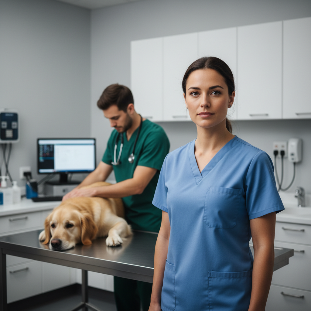
Once you’re at the veterinary clinic, your careful home documentation will provide a valuable starting point. The veterinarian will perform their own physical examination and then recommend a diagnostic plan to determine the nature of the lump. Understanding these common procedures can help alleviate your anxiety about the visit.
Physical Examination and History
Your vet will start by asking questions about when you found the lump, how it has changed, and your dog’s overall health. They will then perform a thorough physical exam, palpating the lump to assess its characteristics and checking the rest of your dog for other abnormalities, including the status of the lymph nodes.
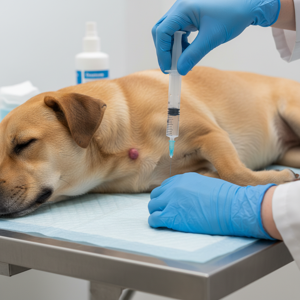
Fine Needle Aspiration (FNA)
This is often the first diagnostic step. It is a simple, minimally invasive, and quick procedure that can often be done in the exam room without sedation. The vet will insert a small, sterile needle into the mass to collect a sample of cells. These cells are then placed on a microscope slide, stained, and examined. An FNA can often differentiate a simple cyst or fatty lipoma from something more suspicious that requires further investigation. However, it’s not always 100% definitive, as the small sample may not be representative of the entire tumor.
Biopsy
If the FNA is inconclusive or suggests malignancy, a biopsy is the gold standard for diagnosis. This involves taking a larger tissue sample for analysis by a pathologist. There are several types:
- Incisional Biopsy: A small piece of the tumor is surgically removed. This is done when the tumor is very large or in a difficult location, and the goal is to get a definitive diagnosis before planning a more extensive removal surgery.
- Excisional Biopsy: The entire lump is surgically removed with ‘margins’ of surrounding healthy tissue. This procedure is both diagnostic (the whole tumor is sent for analysis) and often therapeutic (the tumor is gone).
Biopsies are surgical procedures that require anesthesia. The pathologist’s report will identify the exact type of tumor, state whether it is benign or malignant, and determine if the ‘margins’ are clean, meaning no cancer cells were found at the edge of the removed tissue.
Additional Diagnostics
If cancer is suspected or confirmed, your veterinarian may recommend further tests to check for spread (metastasis). These can include:
- Bloodwork: To assess your dog’s overall organ function and general health.
- Chest X-rays: To check if the cancer has spread to the lungs, a common site for metastasis.
- Abdominal Ultrasound: To examine the abdominal organs for any signs of spread.
The diagnostic process is a systematic way to get a definitive answer. Each step provides more information, allowing your veterinarian to recommend the best possible treatment plan for your beloved companion.
Conclusion
Navigating the discovery of a lump on your dog is a journey from initial fear to informed action. By incorporating a regular, monthly ‘lump check’ into your pet care routine, you become the first line of defense in their health. Remember the key principles: observe, measure, and document. Your detailed notes and ‘lump map’ are not just for your peace of mind; they are critical data that will help your veterinarian make the most accurate assessment.
While this guide provides the tools to conduct a thorough preliminary check, it cannot replace the trained eye and diagnostic capabilities of a veterinary professional. Never hesitate to seek their expertise. Any new lump, or any existing lump that changes, warrants a professional opinion. Your vigilance, combined with professional veterinary care, forms a powerful partnership. By working together, you can ensure your dog receives the best possible care, facing any health challenges with confidence and a clear plan of action.